Free Download: Pelvic Ultrasonography Poster
Get your Free Download: Pelvic Ultrasonography Poster today!
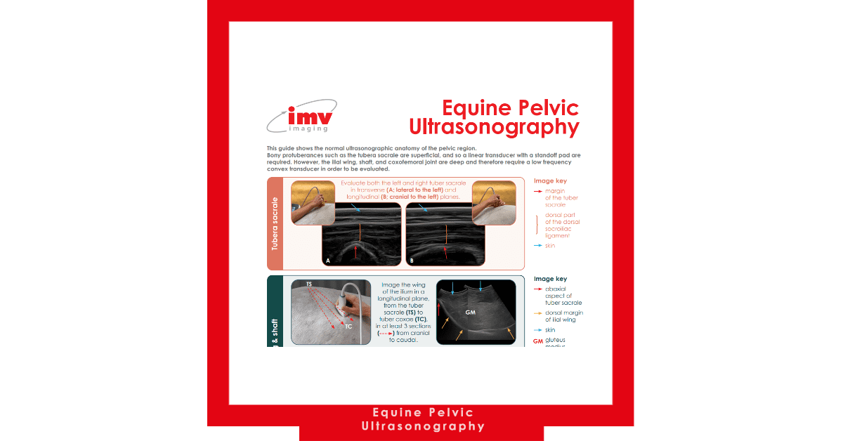
Designed for veterinary professionals, our poster focuses on the tubera sacrale, the ilial wing, shaft, and coxofemoral joint.
Pelvic ultrasonography is a vital diagnostic tool in veterinary practice, offering a non-invasive and detailed view of the pelvic region. Veterinarians use it to assess reproductive health, detect musculoskeletal issues, and diagnose urinary and gastrointestinal conditions in animals, ensuring comprehensive care and accurate diagnoses.

In equine reproduction, pelvic ultrasonography is invaluable. Equine veterinarians can use it to assess the mare’s reproductive organs, including the uterus and ovaries, during breeding cycles and pregnancy. It allows for the detection of ovarian abnormalities, tracking of follicle development, and confirmation of pregnancy. Accurate monitoring of the pregnancy’s progress, including foetal size and well-being, is crucial for ensuring a successful outcome.
Additionally, pelvic ultrasonography aids in diagnosing and managing musculoskeletal issues in horses. It allows veterinarians to visualize the pelvic bones, including the ilium and sacrum, for signs of injury, inflammation, or structural abnormalities. This is particularly important in cases of lameness or performance problems, helping veterinarians tailor treatment plans for the best possible outcomes.
Moreover, equine vets use pelvic ultrasonography to investigate urinary and gastrointestinal conditions. It can reveal issues such as bladder stones, tumours, or gastrointestinal obstructions, allowing for prompt diagnosis and treatment
Get your handy Free Download: Pelvic Ultrasonography poster now to improve your ultrasonography skills and take your equine practice to the next level!


