Case study – 6 yo American Horse with lameness
This 6 year old American Quarter Horse presented with a history of intermittent, severe lameness of the right forelimb of several months duration.
Andrew Hayes BVetMed MRCVS
This 6 year old American Quarter Horse presented with a history of intermittent, severe lameness of the right forelimb of several months duration.
On examination at the yard, the horse was sound in a straight line but resented turning to the right. It was sound when lunged on a soft surface and a hard surface. Under saddle the horse lacked hindlimb impulsion accompanied by a reduction in the hindlimb foot flight arc.
Although bilateral tarso-metatarsal joint blocks improved hind limb action, a right forelimb lameness graded at 1/10 became apparent on the right rein under saddle.

This right fore lameness worsened after the placement of a palmar digital nerve block. It was subsequently graded at 2/10 when the horse was trotted in a straight line on a hard surface. This lameness was not accentuated by forelimb flexion.

After placement of an abaxial sesamoid block the right fore lameness was graded at 3/10 and after a low four point the lameness was graded at 4/10 when the horse was trotted in a straight line.

The lameness got even worse after the placement of a high four point nerve block and was now graded at 5/10.
At this stage the horse was referred to our clinic for further nerve blocking. However upon re-examination two hours later, the horse was sound and despite prolonged lunging and ridden exercise the horse remained sound.
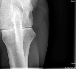
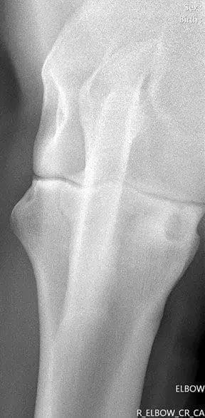
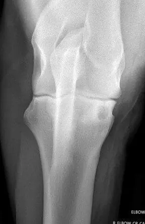
In the absence of any lameness the elbow and shoulder joints of both forelimbs were radiographed.
Craniocaudal views of the right elbow revealed the presence of a well defined osseous cyst-like lesion in the proximomedial aspect of the radius which appeared to be communicating with the elbow joint.
Intra-articular medication of the right elbow with methylprednisolone and sodium hyaluronate has resulted in the resolution of the lameness for the last 3 months.

