Evaluation of ovarian and uterine structures in cows
Correct identification and evaluation of ovarian structures can increase the success and profitability of your reproductive management programs.
Correct identification and evaluation of ovarian structures can increase the success and profitability of your reproductive management programs.
You can assess follicles, the corpus luteum, cystic ovarian structures, and the uterus using ultrasound. By combining ultrasound findings with farm records and observations, the reproductive status of each cow can be determined. This allows you to base treatment and management decisions on these findings and can improve fertility rates within the herd. Similarly, cows with a history of poor fertility can be examined for signs of ovarian and/or uterine pathology, which means appropriate treatment can then be initiated.
Examples of the ultrasonographic appearance of ovarian structures
Follicles

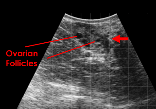
This image shows multiple small ovarian follicles. The fluid filled follicles appear anechoic (black) and are surrounded by the stroma of the ovary (red arrow).

In this image of an ovary, a single, larger follicle can be seen.
Corpus Luteum

The corpus luteum appears as a defined area of hypoechoic tissue within the ovarian stroma.

A central lacuna (fluid-filled cavity) may be seen within a normal corpus luteum.

In this image, two corpora lutea can be seen indicating that a double ovulation has occurred.
Ovarian Cysts
 In this image, a large fluid-filled follicular cyst can be seen. Note the thin wall of the cyst.

This image shows a luteal cyst with a central fluid filled area of 25mm. Note the thicker wall of the cyst containing luteal tissue.
