Udder ultrasonography
Ultrasonography of the udder can provide additional diagnostic information regarding the udder parenchyma.
How to scan
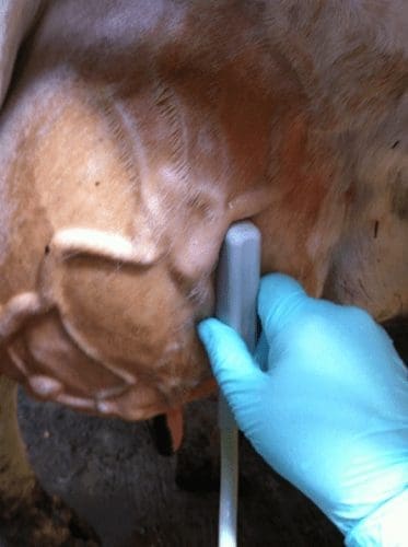
Each quarter of the udder should be examined in a systematic manner. The probe should be positioned in both proximal-distal and cranial-caudal orientations and moved along the surface of each quarter to enable the entire gland to be evaluated (see Figure 1 for technique).

1 – Proximal-distal orientation of probe placement on the udder
Normal anatomy
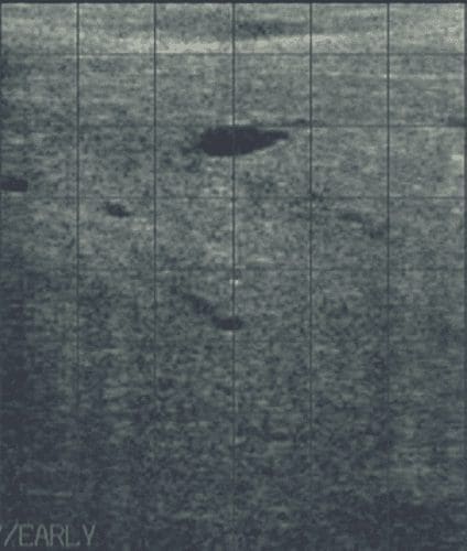
As shown in Figure 2, the parenchyma of the udder should be relatively homogenous with visualization of milk ducts and blood vessels throughout. The degree of distension of the milk ducts will vary in relation to time of milking.

2 – Normal udder parenchyma

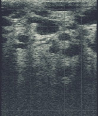
3 -Bovine udder with mastitis caused by Trueperella pyogenes.
Example of what you will see:
Video – Easi-scan normal udder from BCF Technology via Vimeo:
Pathology
Ultrasound may be used to investigate cases of udder enlargement which present without clinical signs of mastitis, such as hematomas, edema, and abscess formation which does not involve the milk producing tissue. In mastitis cases, the ultrasound image of the affected gland may demonstrate abnormalities which have been associated with specific causative agents.
Figure 3 is an ultrasound image of a bovine udder affected with mastitis caused by Trueperella pyogenes. This is characterized by the presence of multiple, hypoechoic regions throughout the parenchyma. Bacterial milk culture confirmed the causative agent.
Why ultrasound?
Ultrasonography of the udder can be a useful adjunct to more commonly used diagnostic tests for the evaluation of udder health such as visual inspection of the udder and milk, udder palpation, somatic cell count evaluation and bacteriological culture of milk samples.