Small animal veterinary case study – Daisy the chihuahua
Case provided by Yvonne McGrotty BVMS CertSAM DipECVIM-CA MRCVS
European Specialist in Veterinary Internal Medicine
RCVS Specialist in Veterinary Internal Medicine
History
Daisy was presented with a 2 day history of tonic clonic seizures and a one week history of altered behaviour (unprovoked aggression).

Two seizures occurred in close succession to each other and each lasted around 60 seconds. Post ictal changes lasted around 90 minutes. Prior to this the owner reported that Daisy was an otherwise normal puppy and was doing well with toilet training. Daisy was being fed James Wellbeloved puppy food and had been vaccinated and dewormed. She was from a litter of 5 puppies and the owner had seen one of the litter mates and both parents and these animals were clinically normal.
Bloods had been collected and revealed reference range albumin and urea (remaining biochemistry was unremarkable). Haematology was unremarkable with no evidence of microcytosis. A mild thrombocytosis was present which may have been physiological. Daisy had a further seizure in the car on her way to Broadleys and the owner had administered around 1.25mg of diazepam rectally.
Physical examination
On clinical exam Daisy was heavily sedated and poorly responsive to stimuli. Heart rate was remarkably slow (100bpm) but pulse quality was good. Thoracic auscultation was unremarkable and no pain was noted on abdominal palpation. Mild hypothermia was present (37.5C). Her cranium did appear fairly domed. A neurological examination could not be performed due to sedative effects of diazepam.
Clinical pathology
A resting ammonia level (after 12hr fast) was within reference range and blood glucose was 5.1mmol/l. Daisy was fed (and she ate with great enthusiasm) and a post-prandial ammonia was also within reference range. A bile acid stimulation test showed adequate liver function.
Diagnostic imaging
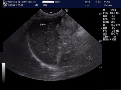
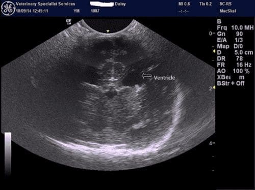
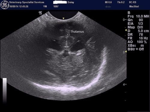
We performed cranial ultrasound via the fontanels. This allowed us a limited view of the brain. An abdominal ultrasound was also performed. The liver was of reasonable size and portal vasculature was identifiable. The kidneys were not enlarged and the rest of the scan was unremarkable.


Daisy has now undergone brain MRI to determine the reason for her seizures.