Small animal veterinary case study – Victor the labrador
History & signalment
Victor – a 9yr MN Labrador was presented with a history of abdominal distension and poor appetite. On examination he was underweight with an enlarged abdomen and abdominal discomfort.
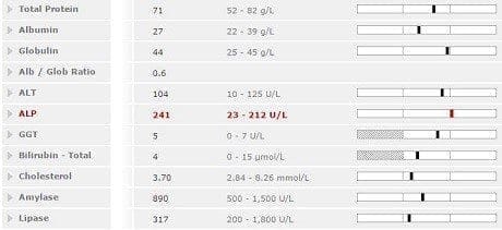
Bloods were fairly unremarkable, showing only a mild increase in ALP. Spec cPL was within reference range.
An abdominal ultrasound was performed. There was no free fluid and no enlarged abdominal lymph nodes. The liver was markedly abnormal. The liver was enlarged with lobes extending well beyond the costal arch. Lobes were rounded and had an irregular, uneven surface. The fat in the cranial abdomen was hyperechoic suggestive of inflammation. A mass was detected within the liver lobe on the right side; this was roughly spherical and parenchymal detail was altered.
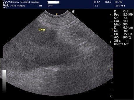
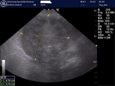
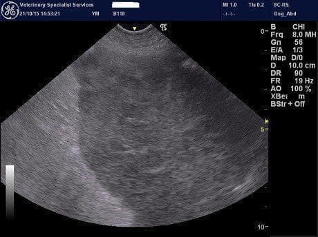
FNAs were obtained from the mass. Cytology was suggestive of an epithelial neoplasm with moderate inflammation. Hepatic carcinoma and biliary carcinoma were differentials in this case.
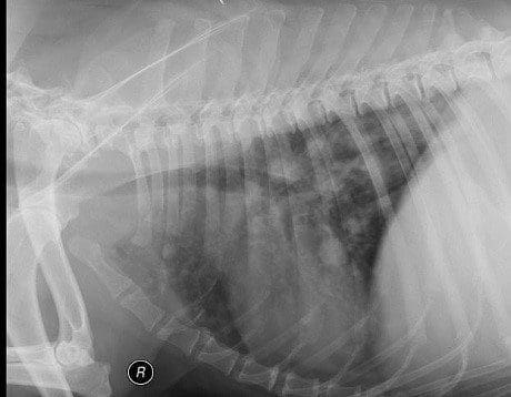
A thoracic radiograph was obtained and sadly revealed multiple nodules throughout the lung fields consistent with metastatic disease. The presence of metastases makes a biliary carcinoma more likely as hepatic carcinomas are generally very slow to spread. Histology would have been necessary to confirm the type of tumour, but was considered pointless given the evidence of spread to the lungs at the time of diagnosis.